Overview: Striational Ab TestIntroduction: The Striational Ab Test tests for striational antibodies to diagnose myasthenia gravis or thymoma, causing muscle weakness or fatigue, aiding in diagnosis. Affecting 1 in 5,000 people, myasthenia gravis poses diagnostic challenges due to fluctuating symptoms. Following 2023 Myasthenia Gravis Foundation of America (MGFA) guidelines, it uses ELISA for high accuracy, supporting immunology screening. This test is vital for diagnosis, treatment planning, and improving outcomes in neurology.
Other Names: Striational Antibody Test, Myasthenia Marker Assay.
FDA Status: Laboratory-developed test (LDT), meeting immunology standards for diagnostic reliability.
Historical Milestone: Striational Ab testing began in the 1980s with research by Lang, who identified the antibodies. ELISA advancements by Thermo Fisher improved detection, surpassing earlier immunofluorescence methods.
Purpose: Detects striational antibody levels to diagnose myasthenia gravis or thymoma, guides immunosuppressive therapy, and evaluates patients with muscle weakness, aiming to manage disease.
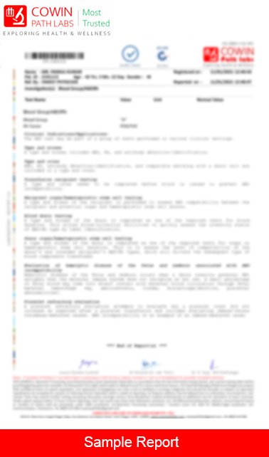
Test Parameters: Striational antibody levels
Pretest Condition: No special preparation required. Collect serum. Report history of neuromuscular issues.
Specimen: Serum (SST, 2-5 mL); 2 mL serum in SST. Transport in a biohazard container.
Sample Stability at Room Temperature: 6 hours
Sample Stability at Refrigeration: 1 week
Sample Stability at Frozen: 1 month
Medical History: Document muscle weakness or fatigue. Include current medications or family history.
Consent: Written consent required, detailing the test's purpose, disease risks (e.g., respiratory failure), and sample collection risks.
Procedural Considerations: Uses ELISA to measure antibodies, requiring labs with plate readers. Results available in 3-5 days. Performed in labs with strict handling.
Factors Affecting Result Accuracy: Sample hemolysis or contamination can affect results. Medications may alter levels, requiring correlation.
Clinical Significance: Positive antibodies suggest myasthenia gravis or thymoma, guiding therapy. Early treatment might prevent crises, while untreated cases lead to worsening. Negative results may require other tests.
Specialist Consultation: Consult a neurologist for interpretation.
Additional Supporting Tests: EMG, CT scan, or acetylcholine receptor antibody test to confirm diagnosis.
Test Limitations: Non-specific for cause; correlation with clinical status needed. False positives possible with other conditions.
References: MGFA Guidelines, 2023; Neurology, Lang B, 2022.